- Stephanix
- Products
-
- News
- Customer service
- Contact

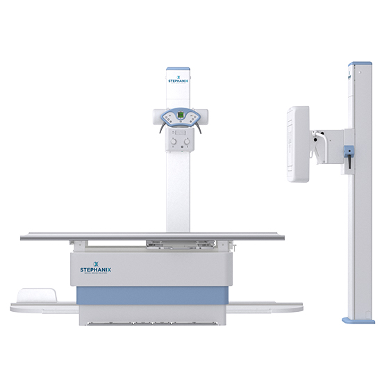
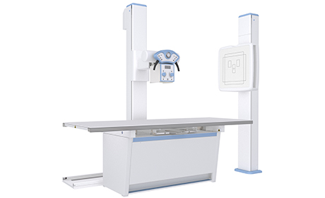
This multipurpose x-room allows you to perform examinations with the image receptor inside the bucky, or outside for direct projections such as weight bearing exams, on stretcher…
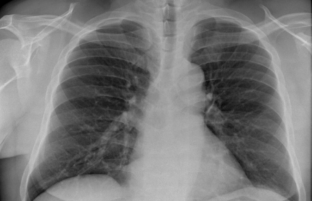
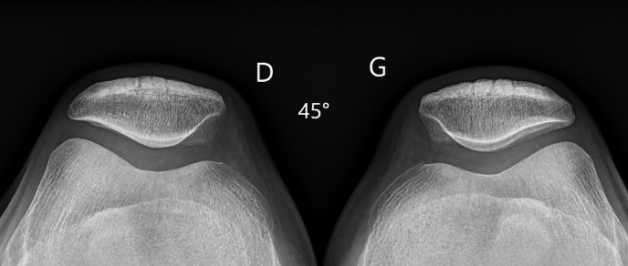
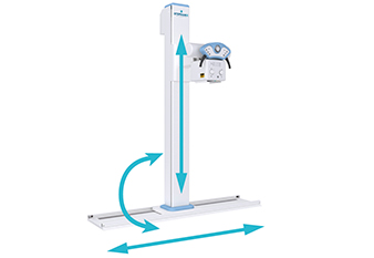
The movements of the C2RS tube holder column, longitudinal on a rail, lateral by a telescopic arm, and vertical, enable optimal positioning to perform many types of examinations.
The rotations of the X-ray unit and the column +/-180° allow acquisition of images in all directions, on table, on wall bucky as well as directly on a stretcher.
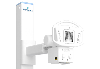
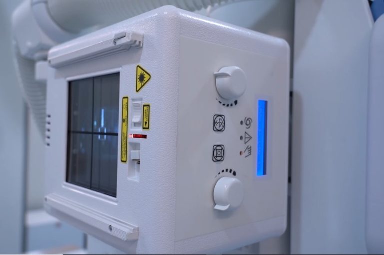
In the automated version, the column is equipped with a control console with touch screen to modify the exposure parameters as well as the adjustment of the stands such as DSI, incidence, collimation, filtration… while remaining close to the patient.
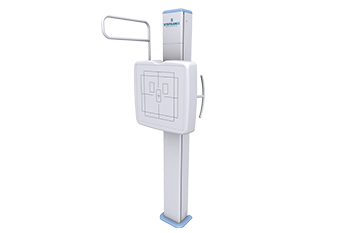
The wall bucky moves vertically to perform whole acquisition of the bones and pulmonary examinations with standing patient.
In auto-tracking or automated configuration, the movements of the tube are synchronized with the movement of the wall bucky.
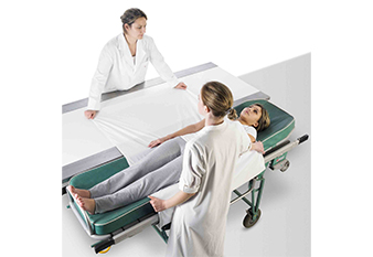
Choose a RAD Series Pro room with an elevating table to adjust the height of the table top to the height of the stretcher for easier patient transfers.
Your RAD Series Pro auto-tracking or automatic x-ray room, with a touch screen on the tube head, has preprogrammed anatomical protocols, acquisition parameters and stand positions.
With the auto-tracking function, the tube of the column follows the vertical movements of the image receiver of the wall bucky or the elevating table.
In the automated version, each stand is automatically positioned, the collimator opens to the image format selected in the protocol, and additional filters are set up according to the examination (pediatrics, etc.).
At any time, you can adjust the tube position or adapt acquisition parameters.
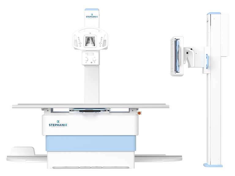
The DR version of your room includes an acquisition console with touch screen interface (in option).
She gives access to patient data entry, choice of anatomical protocols and acquisition parameters, image preview for validation in a few seconds, and image processing before sending to the network.
Each procedure has its own preset parameters such as acquisition parameters, positioning of the stands, collimation, but also additional filtration and post-processing.
The Scatter Radiation Correction option (commonly referred to as Virtual Grid) is performed automatically by software and can be applied to all anatomical regions (at the user’s choice).
It is an image processing algorithm improving the contrast by subtracting the scattered noise.
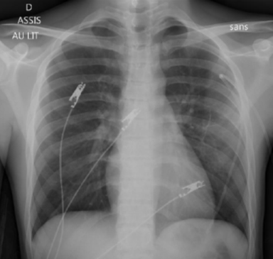
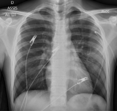
No need to use of physical anti-scatter grids and therefore better ergonomics (no grid to handle) and dose reduction (no grid in the beam), for tabletop or stand examinations as well as for direct or pediatric images.
It also reduces operator and patient exposure due to misalignment and/or artifacts of the focused grid.
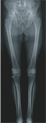
Realization of the long bones radiography
– Acquisition on standing patient with system positioning and pre-programmed adjustment of xray parameters.
– Optimized overlap area for efficient recognition of the pixels used for registration.
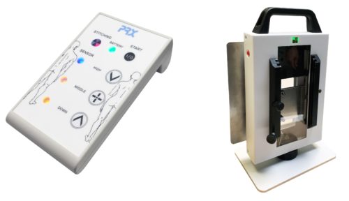
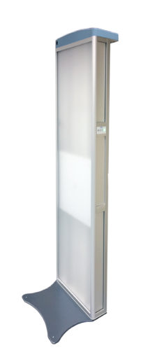
The PRX system is a motorized wall bucky that manages the vertical movement of the DR detector according to the number of images desired to cover the examination area.
The box, which is placed in front of the collimator, controls the radiation by synchronizing asymmetrical collimators with the movement of the DR detector.
The operator sets the bucky to the appropriate height of the first image according to the patient’s size from a control keyboard located on the PRX and starts the automatic acquisition of the examination.
The reconstruction of the examination is then done with the acquisition software which manages the images acquired in auto-trigger mode.
This acquisition mode with the PRX is adaptable to any X-ray source, with a minimum focal distance of 2 meters.
In addition to the optional scatter correction, the dedicated pediatric protocols, the modifiable collimation for each anatomical protocol, the Dose Surface Product (DSP) indicated for each image and the DICOM MPPS and RDSR functions (automatic send of the examination data such as the DSP and acquisition parametrs) are all tools to help you manage the delivered dose as efficient as possible.
Revision date: July 2022

Le contenu de ce site internet est destiné exclusivement aux professionnels de santé.
Je certifie sur l’honneur être un professionnel de santé.