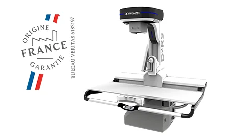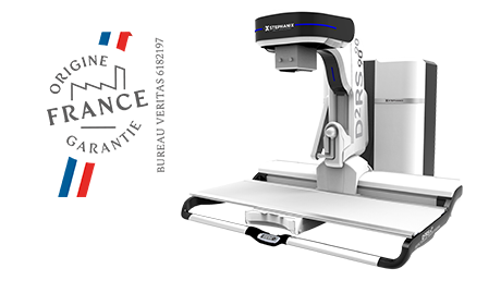


X-ray remote controlled systems that integrate fluoroscopy and radiography a single solution, that meet your expectations: D²RS 90/90 and D²RS:
STEPHANIX has been designing radiology equipment in La Ricamarie, France, for more than 40 years, and remote controlled tables remain our core business. More than 25 years ago, we have integrated a DR flat panel detector in our Evolution x-ray remote controlled table, while fluoroscopy was done with an image intensifier. This demonstrated our commitment to advancing fluoroscopy radiography systems.
More than 10 years ago, STEPHANIX raised the bar by integrating the first PORTABLE DYNAMIC flat panel detector in our D²RS remote controlled table. Inside the bucky the detector works in dynamic and static imaging. Out of the bucky, it is used for direct projections, offering true versatility in fluoroscopy radiography imaging systems.
Once again in 2021 we challenge the status quo with the first Wi-Fi DYNAMIC flat panel detector in the D²RS and D²RS 90/90 x-ray remote controlled tables.
Two configurations answer most clinical and architectural constraints, both compliant with the MDR 2017/745 regulation and supported directly by our teams.
D²RS 90/90
Designed for departments that receive patients with reduced mobility, this remote controlled table drops to a 38 cm (15″) low height and tilts +/- 90°. Transfers become simpler, safer, and considerably less demanding for the radiographer. Full tilt in both directions opens up the widest range of positioning options, from standing chest projections to Trendelenburg examinations, without moving the patient to a second room.
D²RS
Compact by design, with an integrated cabinet and +90°/-25° tilting, the D²RS was conceived for small X-ray rooms where every square metre counts. It suits imaging centres, private practices, and hospital departments that need a versatile fluoroscopy and radiography solution within a constrained footprint. Same dynamic detector philosophy, same image quality, less floor space.
FDA 510(k) cleared · CE-marked MDR 2017/745.
STEPHANIX, French manufacturer and integrator, a talented initiator.
Radiography and fluoroscopy are the two cornerstones of projection imaging. Radiography, commonly called X-ray, uses ionizing radiation to record a static image of internal structures; it excels at bone assessment and fracture detection. Fluoroscopy takes a different approach: a pulsed or continuous X-ray beam generates a live sequence, so the practitioner observes organs and devices in motion rather than frozen in time.
That distinction matters clinically. Watching a contrast bolus travel through the oesophagus, or following a catheter as it advances, is information a single static exposure simply cannot provide.
Our remote controlled tables sit at the heart of these systems. A high-performance dynamic flat panel detector captures real-time images with the clarity required for diagnostic confidence, and the radiologist steers the examination from the control room without repeated entries into the exposure area.
Digital fluoroscopy pairs X-ray generation with modern detector electronics to produce a continuous stream of high-quality digital frames, displayed instantly on the diagnostic console. Swallowing mechanics, joint mobility, contrast dynamics: each becomes observable and recordable. From gastrointestinal studies to interventional guidance, the same room adapts to the day’s schedule, which is precisely what makes these fluoroscopy machines economically defensible for mid-sized departments.
Radio-fluoroscopy equipment serves radiology, gastroenterology, pneumology, urology, and the surgical suite. In radiology, it supplies the precise projections used to identify abnormalities. In interventional settings, it guides instruments in real time, reducing uncertainty during placement. Barium swallow and upper GI series, defecography, hysterosalpingography, myelography, arthrography, catheter and drain positioning, ERCP support: the list of indications remains broad, and it explains why an RF room is often among the most heavily used assets in a department.
Immediacy is the obvious one. The practitioner sees what is happening while it happens, adjusts the projection, and confirms the result without waiting for a processing cycle. Diagnostic uncertainty drops; repeat examinations become rarer.
There is also a workflow argument. One room handles both static and dynamic examinations, so scheduling gains flexibility and capital expenditure is concentrated rather than duplicated. Add pulsed acquisition, last image hold, virtual collimation, and dose area product monitoring, and modern equipment delivers this versatility while keeping exposure aligned with the ALARA principle for both patient and operator.
Digestive tract studies still represent the largest share of activity in most French and European departments. Contrast-enhanced examinations of the oesophagus, stomach, and colon remain the reference indication, closely followed by dynamic musculoskeletal assessment and image-guided procedures.
Beyond diagnosis, fluoroscopy underpins therapeutic guidance: infiltrations, biopsies, device positioning, and drainage placement all benefit from live visualisation. The proportion of interventional work varies considerably from one facility to another, which is why configuration studies matter more than catalogue specifications.
An X-ray tube emits a beam that traverses the patient. Tissues attenuate that beam according to their density; what reaches the detector is a differential signal converted into a digital image several times per second. In legacy installations, an image intensifier and a video chain performed that conversion. Today, a dynamic flat panel detector handles both fluoroscopic sequences and radiographic exposures, with markedly better contrast-to-noise behaviour at equivalent dose.
Automatic exposure control adjusts kV and mA continuously as anatomy moves through the field. Pulsed acquisition, typically selectable between a few frames per second and continuous mode, gives the operator direct control over the dose-versus-motion trade-off. Images are then routed to the diagnostic console and archived through the PACS.
Specification sheets rarely tell the whole story. Detector technology comes first: a dynamic flat panel, ideally portable or Wi-Fi enabled, removes the ceiling imposed by an ageing image intensifier and covers direct projections outside the bucky. Ask about the active surface, the frame rate available in fluoroscopy, and whether the same detector serves both modes.
Then look at the mechanics. Tilt range, minimum table height, patient weight capacity, and source-to-image distance determine which examinations you can actually perform and which patients you can safely accommodate. A 38 cm low position is not a marketing figure; it decides whether a transfer requires one operator or three.
Room dimensions, generator power, dose management features, PACS interoperability, regulatory clearance (FDA registration in the US, MDR 2017/745 in Europe), and the realistic availability of spare parts complete the analysis.
As a French manufacturer and member of the French Healthcare association, STEPHANIX supports installations directly, from the initial layout study to long-term maintenance. Contact our application specialists to review your room constraints and clinical workload; the configuration proposal follows from there.
Fluoroscopy equipment spans a wide price range, whether you are buying in the United States, the United Kingdom, or elsewhere, and the figure depends far more on the category and configuration than on the currency or the brand alone. A refurbished mobile C-arm for surgical use typically starts around $12,000 / €11,000 and can reach $85,000 / €78,000 depending on size and detector generation, while a new unit from an established manufacturer can run from $50,000 / €46,000 well past $150,000 / €138,000. A complete fixed radiography and fluoroscopy (R/F) room — the category our remote controlled tables belong to — represents a larger capital investment, generally starting around $200,000 / €185,000 for a compact, single-detector configuration. Within that range, detector technology remains the single biggest cost driver: a legacy image intensifier costs less upfront than a dynamic flat panel detector, but the flat panel pays for itself through lower dose, faster throughput, and a longer service life before obsolescence. Financing structure matters as much as the sticker price: service coverage, spare-parts availability, and software update policy determine the real cost of ownership over ten to fifteen years of operation. Our remote controlled tables are FDA-registered for the US market and CE-marked under MDR 2017/745 in Europe, so the same platform can be quoted and installed on either side of the Atlantic without a compliance gap. Because every department’s layout, patient mix, and clinical workload differ, we do not publish a flat price list; our team builds a configuration study for each project and issues a firm quotation in your local currency once room constraints and clinical requirements are known.
Fluoroscopy equipment is generally grouped into three families, distinguished by how the system is built and where the operator stands during the exam. Fixed radiography and fluoroscopy (R/F) rooms are permanent installations built around a tilting table, used for the widest range of studies: digestive tract series, urography, orthopaedic and musculoskeletal work, and image-guided procedures. Our D²RS and D²RS 90/90 remote controlled tables belong to this family. Mobile C-arm systems are compact, wheeled units built around a C-shaped arm carrying the tube and detector; they serve mainly in operating rooms and at the bedside for fracture reduction, pain management, or intraoperative guidance. Cardiovascular and interventional systems form a third, more specialised family, built for angiography and cath-lab procedures that demand very high frame rates and fixed ceiling- or floor-mounted gantries. Within the fixed R/F category, machines are further split by how the exam is controlled: tableside-controlled systems are operated from beside the table, while remote controlled systems let the operator run the full examination from a shielded control room. A last distinction worth knowing before comparing quotes is the detector generation: legacy rooms still rely on an image intensifier, whereas current-generation machines, including ours, use a dynamic flat panel detector for both static and live imaging in a single receptor.
In the US market, tableside-controlled fluoroscopy systems are units where the radiographer or physician operates the exposure and table motion standing directly beside the patient, typically with the X-ray tube mounted under the table. This configuration suits quick, single-operator studies and keeps the practitioner in physical contact with the patient throughout the exam. Remote controlled fluoroscopy systems take the opposite approach: the operator drives the entire examination — table tilt, collimation, exposure, and image acquisition — from a leaded control room, viewing the patient through a window or camera feed. For departments running high patient volumes or handling paediatric, bariatric, or immunocompromised patients who need an assistant physically present, remote control removes the operator from the direct beam entirely, which is why it remains the reference architecture in most European hospitals and a growing number of US imaging centers seeking to reduce cumulative staff dose without limiting throughput. Neither architecture is universally better; the right choice depends on staffing model, patient population, and room layout, which is exactly what our team reviews during a pre-installation configuration study.
To provide the best possible experience, we use technologies such as cookies to store and/or access information on devices. Consenting to these technologies will allow us to process data such as browsing behavior or unique identifiers on this site. Not consenting or withdrawing your consent may negatively affect certain features and functions.

Le contenu de ce site internet est destiné exclusivement aux professionnels de santé.
Je certifie sur l’honneur être un professionnel de santé.