

Home > Products > Radiography Systems > Digital Radiography Systems

Every radiology department confronts the same challenge: how to deliver fast, reliable diagnostics while protecting patients from unnecessary radiation exposure and maintaining consistent image quality across dozens of daily examinations. The answer depends entirely on how your digital radiography system is designed, integrated, and calibrated to fit your specific clinical workflow. Stephanix builds complete digital x-ray rooms engineered for the realities of modern practice, not hypothetical ideals.
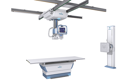
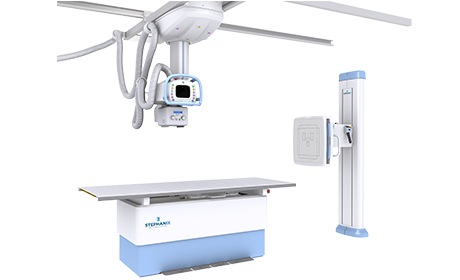
These rooms combine ceiling-suspended or floor-mounted tube supports, fixed or tilting wall Buckys, and fixed or elevating examination tables. Every component is selected to match your clinical volume and physical constraints. The result: imaging environments where technologists work efficiently, patients are positioned comfortably, and diagnostic image quality stays consistent from one exam to the next.


CE-marked.
Selecting the right digital radiography system starts not with spreadsheets but with understanding how your department actually works. How many patients do you scan daily? Which examinations dominate your schedule? What automation level does your team genuinely need, versus what they can operate effectively? These questions determine whether a system enhances your workflow or creates friction.
Some facilities consolidate two separate rooms into a single versatile digital x-ray room, gaining both floor area and workflow speed. Others require specialized configurations for trauma or surgical imaging. Stephanix rad rooms adapt to these diverse scenarios. You choose between column tubestand solutions for compact installations, ceiling-suspended systems for maximum positioning freedom, and universal units combining versatility with ergonomic precision. Each configuration pairs with digital flat panel detectors, advanced image processing software, and customizable table and Bucky setups.
The goal isn’t fitting your department into a standard product. It’s finding the rad room that fits your department’s clinical reality, budgetary constraints, and growth trajectory.
Digital radiography, abbreviated DR, captures x-ray data using flat panel detectors and converts it directly into digital images without film, cassettes, or chemical processing. The x-ray signal transforms into high-resolution digital form almost instantaneously—typically within five seconds of exposure. This speed fundamentally reshapes clinical workflow.
Technologists verify image quality on screen immediately, eliminating repeat exposures. Clinicians receive diagnostic images faster, accelerating clinical decision-making in emergency departments and high-throughput settings. The digital format means images store electronically, share across networks via PACS, and archive without the physical storage demands of traditional film libraries. Radiologists review images on diagnostic workstations within seconds, not hours.
At the core sits the flat panel detector. Direct conversion detectors use amorphous selenium to transform x-ray photons directly into electrical signals, while indirect conversion detectors convert x-rays into visible light using scintillator materials like cesium iodide (CsI) or gadolinium oxysulfide (GOS) before converting that light into electrical signals. Both approaches deliver superior image resolution and dose efficiency compared to older computed radiography plates—typically achieving 2 to 3 times greater dose efficiency than CR, and up to 10 times the dose reduction compared to conventional film, depending on the examination and equipment configuration.
Advanced flat panel detector technology forms the foundation of Stephanix digital radiography systems. These detectors deliver high-resolution x-ray images with excellent contrast and fine anatomical detail. Wide dynamic range means a single exposure clearly visualizes both dense bony structures and soft tissues—the hallmark of true DR capability. This reduces repeat imaging and keeps patient radiation dose minimal.
Rapid image preview distinguishes DR from older technologies. Images appear on the acquisition workstation within seconds, allowing technologists to confirm positioning and exposure quality before the patient leaves the room. This immediate feedback loop virtually eliminates wasted examinations and repeat visits, improving both departmental efficiency and patient satisfaction.
Post-acquisition image processing tools enhance diagnostic value. Contrast adjustment, edge enhancement, magnification, and measurement functions allow clinicians to extract maximum information from every exposure. All image management runs through intuitive software interfaces designed to minimize learning curves for new staff and reduce operational errors through familiar workflows.
Modular room configurations complete the package. Ceiling-suspended or floor-mounted tube stands, fixed or tilting wall Buckys, fixed or elevating tables—these mechanical components combine and customize to match your room dimensions, patient flow patterns, and clinical specialization. The flexibility means no two Stephanix DR rooms need be identical, even within the same facility.
Computed radiography and digital radiography both produce digital images, yet their workflows and capabilities diverge significantly. CR uses reusable phosphor imaging plates in cassettes. After x-ray exposure, a technologist physically carries the cassette to a dedicated CR reader, which scans the plate with a laser to extract image data. The plate then erases for reuse. This process mirrors traditional film-based systems, which made CR an accessible entry point toward digital imaging when introduced. The trade-off: additional handling steps, slower image availability, and generally lower spatial resolution than modern DR panels.
DR eliminates the cassette entirely. The flat panel detector integrates into the x-ray room, capturing images directly and transmitting them to the workstation in real time. Nothing to carry, nothing to scan, nothing to erase. Images appear for review in seconds rather than minutes. Multiply this across dozens of examinations daily and the cumulative time savings reshape departmental throughput measurably.
Image quality favors DR decisively. Flat panel detectors deliver higher spatial resolution, better contrast-to-noise ratio, and wider dynamic range than CR plates. Combined with superior dose efficiency, DR provides clearer diagnostic images at lower radiation levels. For high-volume radiology departments, emergency rooms, and imaging centers where speed and precision directly affect patient outcomes, DR has become the reference standard. CR maintains a role in specific contexts, particularly in facilities with lower examination volumes or short-term budget constraints. However, the global trend remains unmistakable: DR adoption accelerates, while CR systems gradually phase out as flat panel technology becomes more accessible and affordable.
A digital x-ray room combines an x-ray generator and tube with a flat panel detector integrated into the table or wall Bucky. When the technologist triggers the exposure, x-rays penetrate the patient and strike the detector directly, which converts the signal into a digital image displayed on the acquisition workstation within seconds. No cassette transport, no film processing, no manual handling of imaging plates. The workflow gains speed and reproducibility at every step.
A complete digital radiography system comprises the x-ray generator and tube, a mechanical tube support (ceiling-suspended, floor-mounted, or column configuration), an examination table, a wall Bucky for upright imaging, a flat panel detector, and an acquisition workstation with image processing and PACS connectivity software. Stephanix configures each component to match the room’s clinical volume and available space, ensuring your system optimizes for your specific operational demands rather than generic specifications.
Ceiling-suspended tubestands offer maximum freedom of movement and suit high-volume departments with varied exam types. The overhead positioning allows rapid repositioning across multiple patient presentations, from upright to horizontal exposures, with minimal technologist repositioning effort. This flexibility proves invaluable in busy general radiography environments and emergency departments handling diverse trauma cases.
Floor-mounted (column) tubestands occupy minimal floor space and cost less than suspended systems. They work well for smaller rooms or lower patient throughput environments where space constraints are more binding than maximum positioning flexibility. Universal units combine both table and wall Bucky positioning in a single compact footprint, ideal for multipurpose clinics with limited space but diverse examination needs. Your choice depends on your actual examination mix, available square footage, and departmental traffic patterns—not on theoretical ideals.
Most digital radiography rooms require a minimum floor area of around 20 to 25 square meters to allow safe patient positioning, equipment movement, and radiation shielding clearance. However, compact configurations such as universal units (U-arms) can fit smaller spaces while retaining full imaging capability. Exact requirements depend on your tube support configuration and table type. Stephanix can assess your available space and recommend the configuration delivering maximum utility within your physical constraints.
Yes. Many analog rooms retrofit with a flat panel detector and digital generator without replacing the entire mechanical structure, which reduces both cost and installation downtime. Stephanix rad rooms such as the Rad Series E+ are specifically designed to support both analog and digital configurations, allowing facilities to migrate to full DR at their own pace. This retrofit capability matters significantly for hospitals managing capital budgets tightly. Rather than replacing an entire installation, upgrading the detector and generator extends your room’s clinical life while gaining DR speed and image quality advantages.
A digital radiography system’s cost depends primarily on tube support type (ceiling-suspended systems exceed floor-mounted or column units), automation level (autotracking versus manual positioning), detector size and technology, and whether you’re building a new room or retrofitting existing infrastructure. Additional variables include image processing sophistication, PACS integration requirements, and site-specific installation demands. Evaluating total cost of ownership—not just initial purchase price—matters. Include staff training, system maintenance, detector replacement cycles, and productivity gains from faster imaging workflows. A lower-cost system creating bottlenecks or requiring frequent repairs ultimately costs more than a higher-priced system delivering reliable, efficient imaging.
Artificial intelligence increasingly influences modern digital radiography systems, not to replace radiologists but to enhance technologist efficiency and image quality. Intelligent image processing algorithms optimize contrast, sharpness, and visualization of specific anatomies automatically during acquisition. Post-processing tools allow radiologists to adjust image parameters digitally without repeat exposures, recovering diagnostic information that might otherwise require additional patient radiation.
Automated positioning assistance reduces technologist effort and positioning errors. Computer vision systems can detect improper patient alignment during pre-exposure positioning, prompting corrections before the exposure, eliminating wasted examinations and reducing repeat visits. Advanced systems integrate automated exposure control with body part recognition, adjusting generator parameters optimally for the specific anatomy being imaged—whether chest, abdomen, or extremities.
These intelligent systems work within Stephanix digital radiography rooms, particularly in the Xtreme Premium configuration. The goal remains consistent: maximize diagnostic value while minimizing technologist time and patient radiation, ultimately improving departmental throughput and clinical outcomes simultaneously.
To provide the best possible experience, we use technologies such as cookies to store and/or access information on devices. Consenting to these technologies will allow us to process data such as browsing behavior or unique identifiers on this site. Not consenting or withdrawing your consent may negatively affect certain features and functions.

Le contenu de ce site internet est destiné exclusivement aux professionnels de santé.
Je certifie sur l’honneur être un professionnel de santé.