- Stephanix
- Products
-
- News
- Customer service
- Contact

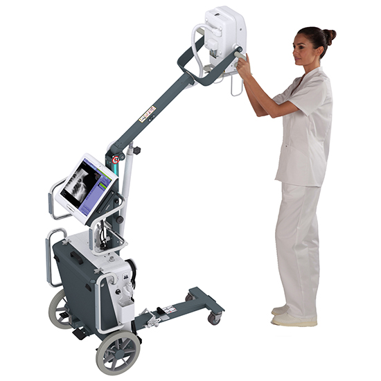
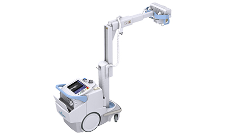
To perform all examinations, imaging in hospitals and nursing homes, at patient’s home … where ambulatory radiography is expected..
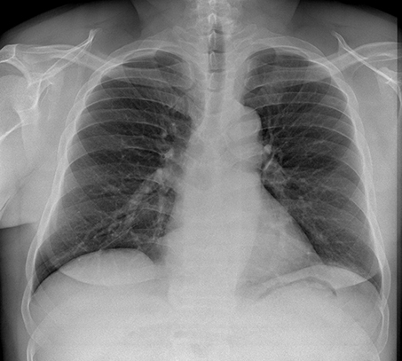
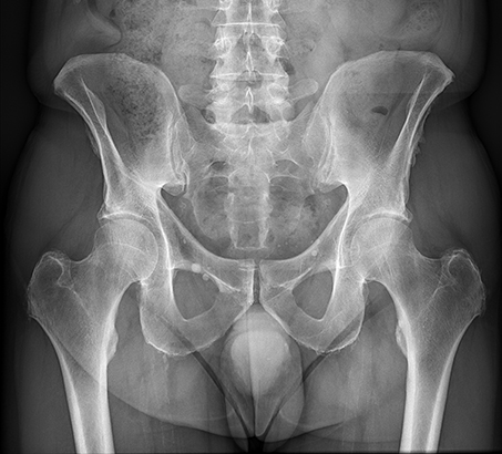
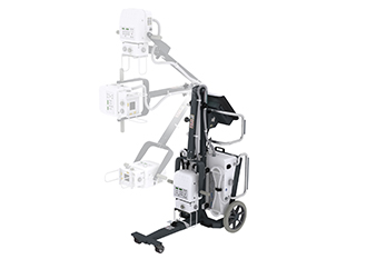
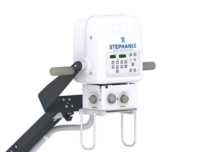
Simply position the mobile by unfolding its arm and turning the tube and collimator to obtain the ideal angle for the examination.
With less than 90 kg combined with its rolling system makes it easy to handle.
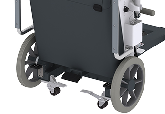
The side wheels are a clever option: press the foot pedal to put them in place, they then help you move the mobile along the bed and adjust its position more easily.
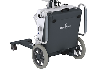
The drawer used for storing the flat panel, and possibly the grid, has a built-in charger (optional) that allows you to charge Flat Panel detector battery.
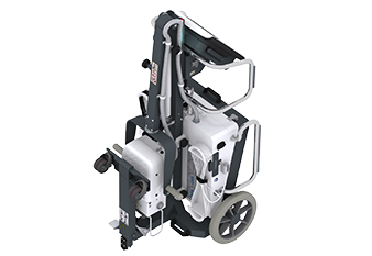
Fold the fork of the front wheels to put the mobile in transport/parking position.
You can then pull it like a golf cart to take to its storage place where it will take less space.
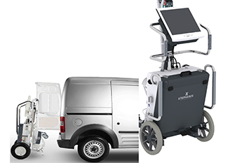
The handles are equipped with plastic reinforcements in order to slide the mobile in your vehicle.
These reinforcements protect the handles from rubbing in your trunk.
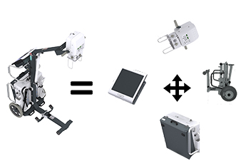
For extreme situations that require special transport conditions (helicopter, dugout …), the mobile can be dismantled in four parts (optional) with usual tools.
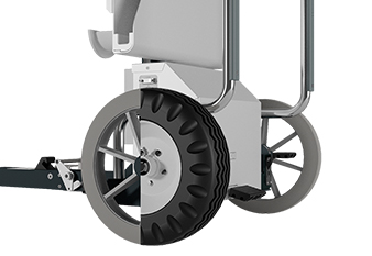
For use in rough areas, during outdoor concerts, on humanitarian or military missions, equip your mobile with pneumatic rear wheels.
They will absorb vibrations from uneven ground.
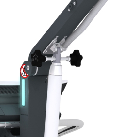
An color coded LED (at the arm junction) indicates the status of the equipment and guide you in preparing for the exam.
Blue: system ready
Green: tube ready
Yellow: exposure
Red: system fault
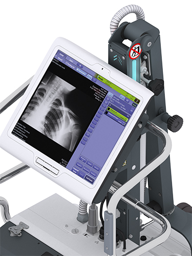
The console of the digital system with DR Detector* has a 17″ touch interface to access the multiple anatomical protocols of the acquisition software.
* This mobile also exists in analog version
Choose the patient from Worklist/Patient list, or manually enter patient data (emergencies) then select the protocol
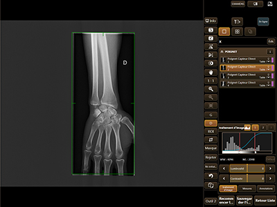
The Scattered Radiation Correction option (usually called Virtual Grid) is performed automatically by software and can be applied to all anatomical regions (as selected by the user).
It is an image processing algorithm that improves the contrast by subtracting the scattered noise.
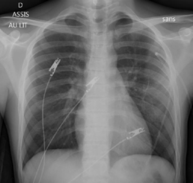
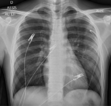
It frees you from the use of physical anti scatter grids and therefore gains in ergonomics (no grid to handle) and provide dose also for pediatric examinations.
It also reduces operator and patient exposure due to misalignment and/or artifacts of the grid.
Optional scatter correction, dedicated pediatric protocols, collimation, Dose Area Product (DAP) indicated for each image ,DICOM MPPS and RDSR functions (automatic sending of exam data such as DAP and acquisition constants) are tools that help you manage the exposure.
Revision date: July 2022

Le contenu de ce site internet est destiné exclusivement aux professionnels de santé.
Je certifie sur l’honneur être un professionnel de santé.